Tag: <span>study design</span>

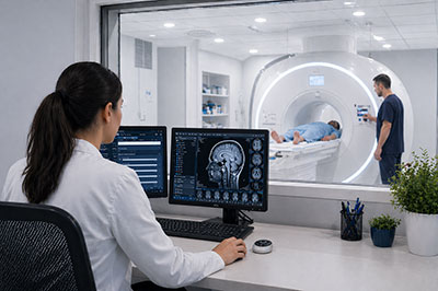
Why We Built Aperis
The thinking behind Aperis — built for how scientists actually interrogate imaging mid-study.
 Gastroenterology
  Oncology
Roughan Sheedy
Managing Director
| Published | February 2025 |
|---|---|
| Category | Platform & Technology |
| Therapy Areas | Gastroenterology, Oncology |
| Read Time | <1 minute |
Related articles

Overview
This article shares perspective from the Fidēs team on early-phase imaging. Replace this placeholder with the full article body — the content is fully editable as blocks.
What this means in practice
Practical implications for sponsors designing imaging into early-phase programs.
The Fidēs approach
How Fidēs embeds imaging into scientific decision-making from protocol design onwards.
The thinking behind Aperis — built for how scientists actually interrogate imaging mid-study.
Read more
Functional cardiac motion as an imaging endpoint in early cardiovascular development.
Read more
The Fidēs team will be at ECCO 2025 in Stockholm — book a meeting to talk early-phase GI imaging.
Read more
The decision problem at the heart of IBD trials, and how imaging can de-risk it earlier.
Read more
Rethinking response assessment in oncology with additive, mechanistically relevant imaging endpoints.
Read more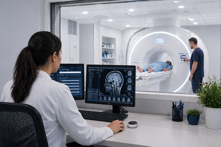
How Aperis gives sponsors continuous visibility of imaging data throughout a Phase I study.
Read more
Why motion and motility are powerful, under-used imaging endpoints in early-phase trials.
Read more
Fidēs Imaging opens its doors as a science-first imaging Core Lab focused on early-phase clinical development.
Read moreBeyond RECIST: Rethinking Imaging Endpoints in Oncology
Rethinking response assessment in oncology with additive, mechanistically relevant imaging endpoints.
Oncology Roughan Sheedy
Managing Director
| Published | January 2025 |
|---|---|
| Category | Science & Research |
| Therapy Areas | Oncology |
| Read Time | 6 minutes |
Related articles

RECIST 1.1 is one of the most widely used frameworks in oncology drug development. Introduced in 2009 as a standardised method for measuring tumour response on CT and MRI, it has provided the field with a common language for reporting and comparing results across programs. That standardisation has genuine value — it is not what this article is arguing against.
What it is arguing is that in Phase I and Phase II programs — where the primary scientific question is often not “did the tumour shrink?” but “is the mechanism doing what we think it is doing?” — RECIST alone frequently fails to capture the biological signal that matters most. And in early development, where go / no-go decisions are made with limited data and high uncertainty, that gap has real consequences.
What RECIST measures — and what it does not
RECIST is a structural measurement framework. It assesses the longest diameter of target lesions on a two-dimensional slice, compares that measurement at successive timepoints, and classifies response as complete response (CR), partial response (PR), stable disease (SD) or progressive disease (PD). It is reproducible, well-understood by regulators and useful for late-phase registration trials where the question is efficacy confirmation.
But it was not designed for the kinds of questions that dominate early development. Specifically, it does not address:
- Functional changes in tumour tissue — such as perfusion, cellularity or metabolic activity — that precede measurable structural changes by weeks or months
- The impact of organ movement on measurement reproducibility — respiratory motion alone introduces systematic error into volumetric lesion assessment that RECIST does not account for
- Secondary biological effects of treatment on surrounding tissue or adjacent organ systems
- Novel or non-standard imaging signals relevant to the specific mechanism of action being studied
For a Phase III confirmatory trial in a well-characterised indication, these limitations are manageable. For a Phase I program exploring a novel mechanism in a patient population where every data point matters, they represent a significant scientific constraint.
The movement problem
One specific limitation deserves more attention than it typically receives: the impact of organ and tissue movement on imaging measurement accuracy.
In thoracic and abdominal oncology programs, respiratory motion during MRI or CT acquisition creates consistent measurement artefacts. A tumour measured at a specific point in the respiratory cycle will appear different in size to the same tumour measured at a different point — not because the tumour has changed, but because the organ has moved. RECIST does not correct for this. It treats each measurement as a static observation.
The difference between a partial response and stable disease on RECIST can, in some cases, be explained entirely by respiratory motion artefact — not by any real change in tumour biology.
This matters at any trial phase, but it matters most in Phase I, where small apparent changes in lesion size can have an outsized influence on dose escalation decisions and early go / no-go signals. The noise introduced by uncorrected motion is not trivial — and it is entirely addressable with the right analytical approach.
Motion correction in practice
Addressing this does not require changing acquisition protocols at site. In most thoracic and abdominal MRI programs, cine-MRI sequences — which capture images across the full motion cycle — are already part of the standard acquisition. The data is present. What is missing is the analytical pipeline to extract it.
Fidēs applies Motilent’s validated GIQuant motion analysis pipeline — developed originally for quantitative GI motility assessment — as the basis for motion-corrected volumetric analysis in oncology programs. The result is a more reproducible lesion volume measurement with significantly reduced inter-timepoint variability that cannot be attributed to real biological change.
Beyond volumetrics: functional imaging endpoints
Even with motion correction, structural volumetrics capture only one dimension of tumour response. In programs targeting novel mechanisms — particularly immune-oncology, anti-angiogenic or metabolic approaches — the earliest detectable signal is often functional rather than structural.
Functional imaging endpoints that complement RECIST in early-phase programs include:
Diffusion-weighted MRI (DWI):
Sensitive to changes in tissue cellularity and water diffusion, which reflect cell death or proliferation before measurable volume changes occur
Dynamic contrast-enhanced MRI (DCE-MRI):
Captures tumour vascular perfusion and permeability — directly relevant to anti-angiogenic and vascular-disrupting agents
PET-CT with FDG or novel tracers:
Metabolic imaging that can detect functional response within days of treatment initiation, well ahead of RECIST-measurable change
Intravoxel incoherent motion (IVIM):
Separates perfusion from diffusion effects within a single MRI acquisition, providing a more complete picture of the tumour microenvironment
None of these replace RECIST for registration purposes. All of them provide earlier and more mechanistically informative signals in early development — which is precisely where the scientific and operational cost of missing a signal is highest.
A comparison of endpoint frameworks by development phase
The table below summarises how common imaging endpoint frameworks map to the scientific questions that are most relevant at different stages of development — and where the gaps are most consequential.
| Framework | Primary Signal | Detects Functional Change | Motion-Corrected | Best Suited To |
|---|---|---|---|---|
| RECIST 1.1 | Structural – lesion diameter | No | No | Phase III / registration |
| iRECIST | Structural – immune response-adjusted | No | No | Phase II / immuno-oncology |
| DWI / ADC | Cellular – diffusion and cellularity | Yes | Partial | Phase I–II / all mechanisms |
| DCE-MRI | Vascular – perfusion and permeability | Yes | Yes | Phase I–II / anti-angiogenic |
| FDG-PET | Metabolic – glucose uptake | Yes | Yes | Phase I–II / metabolic agents |
| Motion-corrected volumetrics | Structural – motion-adjusted lesion volume | No | Yes | Phase I–II / abdominal / thoracic |
What this means in practice: a note on study design
The implication for sponsors is not that standard endpoints should be abandoned — it is that they should be supplemented, deliberately and early, with additive endpoints that are aligned to the specific mechanism being studied and the specific questions that need to be answered before the program moves forward.
Study design considerations
Incorporating additive imaging endpoints at the Phase I stage requires decisions at the protocol level that cannot easily be retrofitted later. The key questions to address during study design are:
- Which functional or motion-based signals are most likely to be sensitive to the mechanism of action — and at what timepoint are they most informative?
- What acquisition parameters are needed to support the additive analysis — and are they compatible with standard site capabilities in the intended geography?
- How will additive endpoint data be integrated with standard RECIST reads in the reporting structure — and what is the regulatory strategy for using it to support go / no-go recommendations?
Video: How Fidēs integrates additive imaging endpoints alongside standard RECIST reads in Phase I oncology programs. Duration approx. 3–4 minutes. To be recorded with the Fidēs scientific team.
Video: How Fidēs integrates additive imaging endpoints alongside standard RECIST reads in Phase I oncology programs. Duration approx. 3–4 minutes. To be recorded with the Fidēs scientific team.
The Fidēs approach
Fidēs was built to support exactly this kind of work — programs where the standard endpoint framework is necessary but not sufficient, and where the value of imaging lies in what it can tell you about biology while there is still time to act on it.
Our study design process starts with the scientific question, not the endpoint list. We work with sponsors’ translational and clinical teams to identify which imaging signals are most mechanistically relevant, which acquisition approaches are feasible within the site network, and how the data will be reviewed and interpreted in real time through Aperis.
The thinking behind Aperis — built for how scientists actually interrogate imaging mid-study.
Read more
Functional cardiac motion as an imaging endpoint in early cardiovascular development.
Read more
The Fidēs team will be at ECCO 2025 in Stockholm — book a meeting to talk early-phase GI imaging.
Read more
The decision problem at the heart of IBD trials, and how imaging can de-risk it earlier.
Read more
Rethinking response assessment in oncology with additive, mechanistically relevant imaging endpoints.
Read more
How Aperis gives sponsors continuous visibility of imaging data throughout a Phase I study.
Read more
Why motion and motility are powerful, under-used imaging endpoints in early-phase trials.
Read more
Fidēs Imaging opens its doors as a science-first imaging Core Lab focused on early-phase clinical development.
Read more